LMORT — Lateral Meniscus Oblique Radial Tears
Anatomy and Biomechanics
A meniscus is a fibrocartilage wedge shaped pad that absorbs shock and stabilizes the joint. Each knee has a medial and a lateral meniscus that rest between the femur and the tibia. The lateral meniscus differs from the medial meniscus by being more circular and more mobile which makes it susceptible to injury in ACL tears.
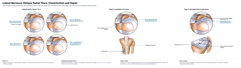
Lateral meniscus oblique radial tears are defined by an oblique radial tear in the posterior horn aspect of the lateral meniscus, measured by its distance from the posterior root. A radial tear cuts the circumferential (hoop) fibers, so the meniscus can no longer resist hoop stress and extrudes from the joint [3]. This can cause major problems for knee stability and long term joint health if left unaddressed.
Presentation
History
Lateral meniscus oblique radial tears are strongly associated with ACL-deficient knees but are not exclusive to them. They mostly affect people under the age of 30, especially men with a higher BMI [5]. Due to the mechanics of the knee, during an ACL tear, the lateral meniscus is often injured, resulting in loss of knee stability. LMORTs present with an acute ACL tear with features of a meniscus tear.
Physical Exam
Patients tend to have generalized posterior lateral pain, but this can be absent. It's important to realize that many timesThere is no overt physical sign of a lateral meniscus root tear, and in some cases, these can even be missed during a routine arthroscopic exam.Because these typically occur with ACL injury the Lachman test, anterior drawer test, and pivot shift tests are the common physical exam maneuvers for assessing the integrity of the ACL [4]. In addition, classic test for the meniscus integrity by performing the McMurray test, the Apley compression test, and the medial-lateral grind test, But again, these will commonly not be present or will be difficult to perform, given the associated injuries in the knee. The Lachman is the most useful single ACL test, while the individual meniscal tests are fairly weak on their own, and a combined exam does better. The most important thing is to be suspicious and closely evaluate for these on the MRI and during the arthroscopic exam.
In some cases, we find patients with persistent posterolateral pain after having a prior ACL reconstruction or partial meniscectomy, which has now weakened through the partial LMORT tear. And in these cases, the patients will present remotely with continued pain a year or more after ACL surgery.
X-Rays
X-rays of your knee will allow your provider to look for fractures, arthritis, alignment issues, and injuries. In acute injuries, these are typically normal, but in chronic cases, there can be narrowing of the lateral joint space. This can compromise results and make a late repair of a lateral meniscal oblique radial tear either more complicated or even contraindicated.
MRI and Joint Arthroscopy
The surgeon will likely order an MRI to evaluate the soft tissues of the knee. It's important to realize that this is separate from the X-rays, but both are needed and helpful. An MRI’s sensitivity with LMORTs varies, especially if there is also an ACL tear present. So diagnosis will combine MRI, the exam, and what is seen in the surgery. Arthroscopy is the definitive test and many LMORTs are first found in ACL surgery. Fellow surgeons up to date with the current literature will be on the lookout for these.
Treatment
LMORTs are classified into 4 different types that depend on the severity of the tear.
- Type 1: partial tear within 10mm of the root
- Type 2: complete tear within 10mm of the root
- Type 3: partial tear more than 10mm from the root
- Type 4: complete tear more than 10mm from the root [5]
The decision to repair depends mainly on how much of the meniscus is torn. In general, if more than about 50% of the radial fibers are disrupted, the tear should be repaired, because the meniscus can no longer hold hoop stress [1,2]. Smaller partial tears may be left alone.
Surgery
Repair is usually performed at the same time as the ACL reconstruction. Tears near the root (types 1 - 2) are typically repaired by reattaching the root to the bone, while tears farther from the root (types 3 - 4) are fixed with a side-to-side repair using spanning sutures [3]. It is important to take care in avoiding the posteriorly located popliteal neurovascular bundle. Repair is preferred over removing the torn piece, because removal does not restore the hoop fibers. The meniscus stays extruded, which puts more force on the ACL graft and the rest of the knee cartilage [1,2].
Rehabilitation
How and when you get back to your regular activities will depend on a number of factors: the type and location of your tear, the repair technique, whether and how your ACL was reconstructed.
What to expect:
- Crutches and weight: Plan on using crutches with little to no weight on the leg for about 4-6 weeks.
- Brace: You will wear a hinged knee brace that is locked straight for about the first 6 weeks.
- Bending: Bending is usually limited to 90 degrees for the first 4-6 weeks. Avoid deep squats, kneeling, or lifting for about 4 months.
- Physical Therapy: PT is recommended about 2-3 times a week along with a home exercise program.
- Return to sports: Anticipate about 6-12 months before returning to sports.
Dr. Gilmer’s take: Root repairs can be difficult to identify preoperatively on MRI, but the literature is leaning towards repairing more of them. I prefer to perform root repairs when near the root and will do side-to-sides with all inside meniscal repair implants for partial tears. My tendency is to repair or reinforce in almost all cases.
If a full root repair, as with a complete injury, then will remain toe-touch weight-bearing for 4 weeks, with range of motion limited to 90°, then progressing to full gait by around 6 weeks. With partial or side-to-side repairs where some of the fibers are intact, we may perform a standard meniscal repair protocol instead. However, because the consequences of high-grade radial tears and root tears are significant I tend to be more conservative with this protocol.
It's important to realize that the lateral meniscus, in addition to dispersing forces and preserving the joint, also contributes stability, prevents the pivot shift phenomenon of the knee, and functions as a secondary stabilizer. This is an additional reason to repair them at the time of ACL surgery.
Risks
This surgery is complex and there are some specific complications that can occur:
- Post-operative fluid in the knee (can require drainage in the office). Swelling more than 4 to 6 months after the surgery suggests ongoing cartilage injury or failure of the repair.
- Continued pain and stiffness due to scarring in the knee, occasionally requiring surgery to restore motion. Though more commonly related to ligament surgery, some stiffness can occur.
- Need for re-operation to address a new meniscal tear or scarring. Even when these repairs do not fail outright, they can have residual laxity or some residual extrusion, and this has been shown in some studies.
- New meniscus tear or articular cartilage injury. Despite successful initial treatment, there can be recurrent injury later.
- Development or progression of knee arthritis. This is the primary concern which repair seeks to avoid, but nonetheless, there can be cartilage damage at the time of injury that takes time to manifest.
- One specific risk to this is that passage of all-inside meniscal repair anchors in this location can be difficult and dangerous because the neurovascular bundle of the knee is very close to the posterior horn of the lateral meniscus.
A more general complication of surgery can also occur and would include:
- Deep venous thrombosis (aka “blood clot” or DVT)
- If you have a history of clots, make sure to tell your surgeon - blood thinners may be used in this subset of patients to prevent clots after surgery.
- Infection (all patients receive antibiotics at the time of surgery to decrease this risk)
- Nerve injury (associated with numbness, weakness, or paralysis). As noted, this is particularly important with this particular procedure because of the proximity to the tissue being sewn.
- Vascular injury or compartment syndrome
- Complications associated with the anesthesia
References
- Smith PA, Bezold WA, Cook CR, et al. Kinematic analysis of lateral meniscal oblique radial tears in the anterior cruciate ligament - deficient knee. Am J Sports Med. 2021;49(14):3898-3905. doi:10.1177/03635465211052521
- Karom J, Peterson C, Cavendish P, et al. Lateral meniscus oblique radial tears: a review of diagnostic considerations, biomechanical impact, and treatment outcomes. JBJS Rev. 2025;13(8):e25.00102. doi:10.2106/JBJS.RVW.25.00102
- Herring SA, Kibler WB, Putukian M, et al. Initial assessment and management of select musculoskeletal injuries: a team physician consensus statement. Med Sci Sports Exerc. 2024;56(3):385-401. doi:10.1249/MSS.0000000000003324
- Solomon DH, Simel DL, Bates DW, Katz JN, Schaffer JL. The rational clinical examination. Does this patient have a torn meniscus or ligament of the knee? Value of the physical examination. JAMA. 2001;286(13):1610-1620. doi:10.1001/jama.286.13.1610
- Krych AJ, LaPrade MD, Cook CS, et al. Lateral meniscal oblique radial tears are common with ACL injury: a classification system based on arthroscopic tear patterns in 600 consecutive patients. Orthop J Sports Med. 2020;8(5):2325967120921737. doi:10.1177/2325967120921737
- O’Donnell K, Freedman KB, Tjoumakaris FP. Rehabilitation protocols after isolated meniscal repair: a systematic review. Am J Sports Med. 2017;45(7):1687-1697. doi:10.1177/0363546516667578