Painful Osgood-Schlatter Disease in Adults: Treatment Overview and a Surgical Example
Kathleen L. Hopps, MS2, Brian Gilmer, MD
Osgood-Schlatter disease (OSD), also referred to as tibial insertion disease, is a traction apophysitis of the tibial tuberosity and the most common osteochondrosis of the lower limb in sport-participating children and adolescents [1]. Repetitive tensile loading through the quadriceps and patellar tendon at its insertion on the immature tuberosity produces localized pain, swelling, and tenderness, typically aggravated by kneeling and activity [1]. Most cases resolve with skeletal maturity, but a subset persists into adulthood, often with an ununited ossicle [2], [3]. This work summarizes the presentation, diagnosis, and treatment of painful OSD, with emphasis on the adult presentation, and then describes one surgical example.
Epidemiology and Presentation
Onset coincides with development of the secondary ossification center of the tibial tuberosity, and symptoms typically first appear between ages 8 and 12 in girls and 12 and 15 in boys [1]. The tibial tubercle is cartilaginous before about age 11, the apophysis forms at about 11 to 14 years, it fuses with the tibial epiphysis at about 14 to 18, and the proximal tibia is fully fused after about age 18. This sequence accounts for the usual resolution of symptoms with skeletal maturity. One ultrasonographic study found a peak prevalence of symptomatic OSD at approximately age 12 in both sexes, with an earlier secondary peak in girls (ages 9 to 10) and a later one in boys (age 14) [4]. Approximately 1 in 10 athletic adolescents are affected, with higher rates in sports involving jumping, kicking, and rapid changes of direction, such as soccer, basketball, gymnastics, and volleyball. Involvement may be unilateral or bilateral, with roughly 20 to 30% of cases bilateral [1].
Course and Persistence into Adulthood
OSD is generally self-limiting, and reported rates of resolution with conservative management range from about 80% to 90% [1], [5]. A minority, an estimated 10%, remain symptomatic into adulthood. Follow-up studies report persistent pain in a substantial proportion, including nearly 40% of patients in one cohort and a median symptom duration of 90 months in another [1]. Adult presentations frequently involve an ununited ossicle at or posterior to the patellar tendon insertion, at times accompanied by a prominent tubercle that can irritate the tendon [3]. Refractory cases are managed operatively, with ossicle excision considered in skeletally mature patients with persistent symptoms [1], [2].
Diagnosis and Imaging
Diagnosis is predominantly clinical, based on history and focal tenderness and swelling over the tibial tuberosity. Typical symptoms are anterior knee pain exacerbated by kneeling, and examination shows an enlarged, tender tibial tubercle with pain on resisted knee extension. Imaging (radiography, ultrasonography, or MRI) can help to exclude fracture, tumor, infection, tendinitis, and Hoffa’s disease and to characterize disease stage [1]. The recommended radiograph view is a lateral knee x-ray to assess tuberosity irregularity, elevation, lucency, or fragmentation in advanced disease [1]. Ultrasonography is inexpensive and can show fragmentation of the ossification center, tendon abnormalities, edema, and reactive bursitis. MRI is the most sensitive modality and has been used to stage disease, with the terminal stage characterized by complete ossicle separation and tendon thickening. MRI may also show soft tissue swelling, thickening and edema of the inferior patellar tendon, and fragmentation and irregularity of the ossification center. However, MRI is not essential, as the diagnosis can be made from history, tender swelling, and radiographs alone.
Dr. Gilmer's take: I am more commonly obtaining an MRI if we are:
- Looking for a location to place a platelet-rich plasma injection at the area of the worst tendinopathy, although sometimes this can be detected on ultrasound alone
- Considering surgical treatment Is the primary reason I obtain an MRI
Risk Factors and Complications
Proposed contributors include the adolescent growth spurt, jumping, sprinting, and kicking activities, high or rapidly escalating training loads, quadriceps and hamstring tightness, limited ankle dorsiflexion, and anatomic variables such as posterior tibial slope and patellar position, although the etiology is likely multifactorial and not fully established [1]. Chronic disease has been associated with bone fragmentation and migration, patella alta, reduced knee flexion, pseudarthrosis, and osteochondromatosis [1].
Ossicle Location Guides Management
In persistent symptomatic Osgood-Schlatter disease, the location of residual ossicles may influence surgical management. Ossicles typically lie adjacent to the tibial tubercle beneath the patellar tendon, but larger fragments may be embedded within the tendon or extend into the infrapatellar fat pad or knee joint, potentially contributing to different pain patterns [2]. Distinguishing intratendinous from intra-articular ossicles is important when planning surgical excision, as their location may influence the choice between open and arthroscopic approaches [6].
Dr. Gilmer's take:
If not truly embedded in the tendon, I like an anterior arthroscopic approach, working along the fat pad. In cases where it is more directly attached to the tendon, I will make a small open incision just medialTo the tibial tubercle to allow access to any adhesions and perform a repair of the tendon into its bony bed if necessary with an all-suture anchor.
Impact on Quadriceps Function
Osgood-Schlatter disease may also affect quadriceps function, although direct evidence is limited. Because the patellar tendon transmits quadriceps force to the tibial tubercle, persistent insertional pain may contribute to quadriceps weakness or reduced voluntary muscle activation. In patellar tendinopathy, quadriceps activation (84.4% vs. 97.9% in controls) and knee extension strength (2.41 vs. 3.37 Nm/kg) were lower, with greater deficits observed when pain and structural damage coexisted [7]. This is most likely related to pain inhibition.
Treatment
Non-operative Care
First-line management of Osgood-Schlatter disease is conservative and focuses on symptom control and progressive rehabilitation. Treatment includes activity modification to reduce painful running, jumping, and cutting, with lower-impact alternatives such as swimming or cycling as tolerated.
Cryotherapy and NSAIDs may help relieve pain, while quadriceps, hamstring, calf, and hip strengthening and flexibility exercises can be incorporated into physical therapy. In some cases, protective padding or infrapatellar straps may be used for symptom relief.
A prospective cohort study found an intervention combining activity modification, pain monitoring, progressive strengthening, and a graded return-to-sport program was associated with successful outcomes in 80% of patients at 12 weeks and 90% at 12 months [8].
Extracorporeal shockwave therapy has also been explored, although evidence supporting its routine use remains limited.
Corticosteroid injections are generally discouraged because of concerns regarding tendon damage.
PRP injections have been investigated for persistent symptoms, but evidence remains limited, and PRP is not established as first-line treatment. When performed, this is typically done under ultrasound guidance.
Surgical Treatment
In adults with persistent symptomatic Osgood-Schlatter disease, surgical intervention may be considered when conservative management fails, and residual ossicles or tibial tubercle prominence contribute to ongoing pain and functional limitation. No standardized surgical approach exists, and the available evidence consists primarily of case reports and small case series [2]. Surgical techniques are selected according to ossicle location, patellar tendon involvement, and the presence of associated bony prominence or intra-articular pathology.
- Arthroscopic or bursoscopic debridement and ossicle excision. These approaches are minimally invasive and allow for assessment and treatment of intra-articular pathology and removal of accessible ossicles while limiting disruption of the patellar tendon. A superolateral viewing portal may facilitate visualization of large or deep ossicles, and a burr or punch can be used for excision. Fluoroscopy may assist in confirming complete removal [2], [9], [10].
Dr. Gilmer's take: This allows for rapid mobilization and a faster recovery. Patients are provided crutches for pain control but have no restrictions of range of motion or strengthening. Though, because they are generally limited by pain, they will continue to require some physical therapy and reconditioning generally for 4 to 6 weeks.
- Open ossicle excision. Open excision provides direct access to ossicles embedded within or closely associated with the patellar tendon, allowing careful removal and tendon repair when indicated [3], [6].
Dr. Gilmer's take: As noted above, I like this approach more commonly if there is concern that we would injure the tendon or the ossicle is so large that it simply needs a larger incision for removal.
- Combined arthroscopic debridement and open ossicle resection with patellar tendon repair. This approach combines arthroscopic evaluation and debridement with open removal of residual ossicles. When excision compromises the distal patellar tendon attachment, repair to the tibial tubercle may be performed using suture anchors. Restoration of the tendon’s appropriate position and tension is important to avoid overconstraint [6].
Dr. Gilmer's take: Since this is most commonly the case, I typically perform a combined procedure in most cases. Considering that small ossicles are often less symptomatic and therefore less likely to require surgical management, and larger ossicles are big but do not allow debridement of the posterior tendon or fat pad, overall, a combined approach is ideal in most cases.
- Tibial tubercle reduction osteotomy or tubercleplasty. These procedures may be considered when residual tubercle prominence or a prominent bony beak contributes to patellar tendon impingement [11]. In one adult surgical series, a beak was present in 71% of patients, and osteotomy was performed in 85% [3].
Dr. Gilmer's take: I do not typically find this necessary except in the most rare and unusual of cases. In some unique cases, I excised the tubercle and repaired the tendon with a suture bridge-type construct directly onto the tibial tubercle Base after it has had the residual apophysis removed.
- Other techniques. Additional procedures described for persistent adult OSD include drilling, bone peg insertion, percutaneous tubercle fixation, and closing-wedge osteotomy. These techniques address different underlying anatomical abnormalities and are selected according to individual pathology [6], [12].
Case Example: Arthroscopic Debridement and Open Ossicle Resection
To illustrate the surgical management of persistent symptomatic Osgood-Schlatter disease in adults, we present the case of an adult female elite figure skater with a history of right anterior knee pain. Her training involved prolonged, high-volume jump training with repetitive unilateral landings, predominantly loading the right (dominant) lower extremity. This repetitive loading may have contributed to cumulative stress at the distal patellar tendon and tibial tubercle.
Imaging demonstrated a 1.25 x 1.5 cm ossicle within the distal patellar tendon. Her history was notable for an L5-S1 microdiscectomy four years earlier. Prior to surgery, she underwent extensive non-operative treatment, including nine months of physical therapy, NSAIDs (topical and oral), steroids (oral prednisone), infrapatellar fat pad hydrodissection (5 cc ropivacaine + 5 cc dextrose 5%), many types of Cho-Pat straps, and five months of ECSW therapy. Force plate analysis demonstrated marked asymmetric force distribution, with measurable reduction in peak ground reaction force, rate of force development, and limb symmetry index on the ossicle-affected side. These objective findings correlated with subjective reports of instability, fatigue, and declining jump performance, particularly during landing phases requiring eccentric quadriceps loading.
Given her persistent symptoms despite conservative treatment, she underwent surgery with Dr. Gilmer in April 2026. This procedure included arthroscopy, infrapatellar bursa synovectomy, open excision of the ossicle with patellar tendon debridement, and tendon repair with suture anchor fixation to restore the distal patellar tendon attachment. Arthroscopic evaluation allowed for assessment of intra-articular pathology, while synovectomy addressed the chronic inflammatory burden of the infrapatellar bursa. Suture anchor repair was used to restore the tendon attachment following ossicle excision.
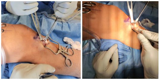
Figure 1. Intra-op Images of the left knee medial aspect show a small open incision and the exposed ossicle.
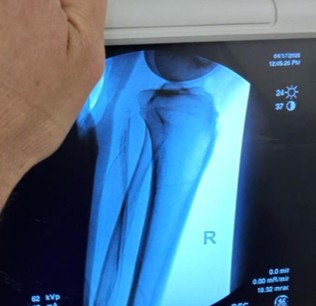
Figure 2. X-ray Fluoroscopic images intraoperatively are used to compare to the preoperative films and ensure complete resection of the painful ossicle. Because it can be surprisingly difficult, I like to routinely confirm complete excision.
This case illustrates the role of a combined arthroscopic and open approach for persistent symptomatic Osgood-Schlatter disease associated with a large ossicle refractory to extensive conservative management. Objective functional testing (such as Force plates) may complement patient-reported symptoms by identifying biomechanical deficits that affect athletic performance. However, as a single case, these findings cannot establish treatment efficacy or predict outcomes in other patients.
Dr. Gilmer's take:
In summary, this patient underwent a combined approach with arthroscopic debridement of the tendon, trephination of the area of primary tendinosis, and a small open excision of the ossicle (with placement of a single suture anchor repairing the tendon into the residual bed to secure it to the remaining united tubercle).The patient proceeded to return to high-level activities, including skating.
References
- F. Corbi et al., “Osgood-Schlatter Disease: Appearance, Diagnosis and Treatment: A Narrative Review,” Healthcare (Basel), vol. 10, no. 6, p. 1011, May 2022, doi: 10.3390/healthcare10061011.
- W. Choi and K. Jung, “Intra-articular Large Ossicle Associated to Osgood-Schlatter Disease,” Cureus, vol. 10, no. 7, p. e3008, 2018, doi: 10.7759/cureus.3008.
- T. F. El-Husseini and A. A. Abdelgawad, “Results of surgical treatment of unresolved Osgood-Schlatter disease in adults,” J Knee Surg, vol. 23, no. 2, pp. 103–107, Jun. 2010, doi: 10.1055/s-0030-1267474.
- Y. Kaneuchi et al., “Bony Maturity of the Tibial Tuberosity With Regard to Age and Sex and Its Relationship to Pathogenesis of Osgood-Schlatter Disease: An Ultrasonographic Study,” Orthop J Sports Med, vol. 6, no. 1, p. 2325967117749184, Jan. 2018, doi: 10.1177/2325967117749184.
- F. Lazko, D. Ananin, A. Petrosyan, M. Awad, M. Panin, and O. Bawareed, “Invasive treatments for Osgood-Schlatter disease (systematic literature review),” Genij Ortopedii, vol. 28, pp. 852–857, Dec. 2022, doi: 10.18019/1028-4427-2022-28-6-852-857.
- F. R. Preuss, R. J. Whalen, W. H. Buchalter, P. Ganokroj, B. T. Provencher, and M. T. Provencher, “Osgood-Schlatter Disease: Ossicle Resection and Patellar Tendon Repair in a Symptomatic Adult,” Arthrosc Tech, vol. 13, no. 11, p. 103110, Jul. 2024, doi: 10.1016/j.eats.2024.103110.
- S. M. Davi, A. S. Lepley, C. R. Denegar, L. J. DiStefan, C. M. Edgar, and L. K. Lepley, “Quadriceps Inhibition After Naturally Occurring Patellar Tendon Damage and Pain.,” Journal of Athletic Training (KnowledgeWorks Global, Ltd), vol. 55, no. 6, pp. 608–614, Jun. 2020, doi: 10.4085/1062-6050-27-19.
- M. S. Rathleff et al., “Activity Modification and Knee Strengthening for Osgood-Schlatter Disease: A Prospective Cohort Study,” Orthop J Sports Med, vol. 8, no. 4, p. 2325967120911106, Apr. 2020, doi: 10.1177/2325967120911106.
- A. K. Alqawlaq and M. Maddah, “Arthroscopic management of refractory Osgood–Schlatter disease: a case series and surgical technique,” J Surg Case Rep, vol. 2025, no. 12, p. rjaf990, Dec. 2025, doi: 10.1093/jscr/rjaf990.
- [ L. I. Zhi-Yao, “Arthroscopic Excision of a Huge Ununited Ossicle Due to Osgood-Schlatter Disease in an Adult Patient,” J Orthop Case Rep, vol. 3, no. 2, pp. 4–7, 2013, doi: 10.13107/jocr.2250-0685.092.
- G. Pagenstert, M. Wurm, S. Gehmert, and C. Egloff, “Reduction Osteotomy of the Prominent Tibial Tubercle After Osgood-Schlatter Disease,” Arthroscopy, vol. 33, no. 8, pp. 1551–1557, Aug. 2017, doi: 10.1016/j.arthro.2017.02.012.
- N. Narayan, P. D. Mitchell, and M. D. Latimer, “Complete resolution of the symptoms of refractory Osgood-Schlatter disease following percutaneous fixation of the tibial tuberosity,” BMJ Case Rep, vol. 2015, p. bcr2014206734, Feb. 2015, doi: 10.1136/bcr-2014-206734.