Kathleen Hopps, MS2, Brian Gilmer, MD
Anatomy and Biomechanics of the Patellar Tendon
The patellar tendon connects the inferior pole of the patella to the tibial tubercle, forming the final link in the knee extensor mechanism. When the quadriceps contracts, force passes through the patella and into this tendon before reaching the tibia, so the tendon absorbs a large share of the load generated when jumping, squatting, or landing. Most of this load concentrates at the two ends of the tendon rather than in the middle, which is why the proximal attachment at the inferior pole of the patella is by far the most common site of pathology [1].
Repetitive jumping and landing place the tendon through repeated cycles of stretch and recoil, and when this loading outpaces the tendon's ability to recover, the tissue begins to change. This is best described by the continuum model of tendon pathology, which breaks the process into three overlapping stages [2].
In the first, reactive stage, the tendon thickens and becomes more cellular as a short-term adaptive response, and this stage is typically reversible with a reduction in load. If loading continues, the tendon enters a state of disrepair, where the collagen matrix begins to break down faster than it can be rebuilt.
With continued overload, the tendon progresses to a degenerative stage, marked by areas of disorganized collagen, abnormal blood vessel growth, and cell death; this stage is far less responsive to simple rest. Many painful tendons are not cleanly in one stage: a small degenerative core is common even in tendons that are still mostly reactive, which helps explain why two patients with a similar pain history can have very different findings on MRI.
Presentation
History
Patellar tendinopathy is most common in athletes whose sport involves repetitive jumping and landing, such as volleyball, basketball, track and field jump events, and soccer, and has been reported in over 40% of some elite jumping populations [3]. Patients typically describe anterior knee pain centered just below the kneecap that is worse with activities which load the tendon quickly, such as jumping, landing, or decelerating, and improves with rest. A useful way to gauge severity is the Blazina classification, originally described for this condition: stage 1 is pain only after activity, stage 2 is pain at the start of activity that fades with warm-up, stage 3 is pain throughout activity and at rest that limits sport, and stage 4 represents a tendon tear [4]. Surgery is generally reserved for patients with Blazina stage III symptoms (pain at rest and during activity, limiting sport) that have persisted for several months despite a structured course of physical therapy, typically at least 3 to 6 months.
Physical Exam
On exam, the tendon is tender to palpation at the inferior pole of the patella, and this tenderness is usually most pronounced with the knee fully extended. One helpful confirmatory test is the Royal London Hospital test: the examiner palpates the tender spot with the knee extended, then repeats palpation with the knee bent to 90 degrees. Because bending the knee pulls the tendon taut and lifts it away from the underlying fat pad, true tendon pain should lessen noticeably in this flexed position; if it does not, another source of anterior knee pain should be considered [5]. Loading tests such as a single-leg decline squat are also useful, both to confirm the diagnosis and to track progress with treatment over time, and many clinicians use the Victorian Institute of Sport Assessment (VISA) score for patellar tendinopathy for the same purpose [6].
Imaging
Plain x-rays are usually normal but are still useful to rule out other causes of anterior knee pain, such as Sinding-Larsen-Johansson disease in a skeletally immature patient or a bony spur at the inferior pole. MRI is the most important study once the diagnosis is suspected clinically. It confirms tendinosis, shows how much of the tendon is involved, and, importantly for deciding on treatment, can show whether the deep fibers of the tendon have begun to peel away from the undersurface of the patella, a finding referred to as delamination [7]. This distinction matters a great deal: a delaminated tendon behaves more like a partial tear than a simple tendinopathy, and tends not to fully resolve with rehabilitation or injections alone [3], [8].
Treatment
Treatment is stepwise, beginning with physical therapy, leukocyte-rich platelet-rich plasma (PRP) injection with needling, MRI to assess for delamination, and then surgical debridement and repair via an arthroscopic approach.
1. Structured physical therapy.
The first and most important treatment is a supervised loading program, usually heavy slow resistance training or eccentric decline-squat exercises, which extensive supporting evidence for patellar tendinopathy [1], [7]. Isometric holds are often introduced early to calm pain before progressing to heavier, more dynamic loading. Given an adequate trial, typically at least 12 weeks, many patients may not require further treatment.
Dr. Gilmer’s take: We often call this eccentric decline a negative: keeping the tendon under load while you slowly enter the squat, emphasizing the descent rather than with coming back up (the focus of concentric exercises, or the more typical way that people do squats). It's not always practical to continue physical therapy for 12 weeks. We usually start with 6 weeks and reevaluate. If there is improvement, then stay the course. However, if there is deterioration or continuing symptoms, we often won't wait a full 12 weeks. The most important thing is not the time spent, but whether the recovery is progressing, even slowly, or is completely stalled.
2. Leukocyte-rich PRP with needling.
If a patient plateaus despite a good effort at therapy, our next step is an ultrasound-guided injection of leukocyte-rich platelet-rich plasma combined with dry needling of the diseased portion of the tendon, continued alongside ongoing loading exercises. One randomized trial found that adding PRP to needling sped up recovery at 12 weeks compared with needling alone, though that advantage had mostly disappeared by 26 weeks [6]. However, other trials comparing different PRP formulations with needling or saline have not shown a clear, lasting benefit from the PRP itself [9]. In practice, needling appears to do much of the work at this stage, and PRP is used to accelerate recovery in the right patient [6], [9].
Dr. Gilmer's take: In practice, I utilize the Stryker Regen-kit system and use a technique of needling under ultrasound guidance, watching the tendon be entered by the needle and then hydrodissected with the platelet-rich plasma as the needle finds and breaks up the areas of most abnormal-appearing tendon.One unfortunate thing about PRP injections is this particular location tends to be very painful, and local anesthetics interfere with degranulation of the platelets. Nonetheless, patients generally can resume light activity, return to work, drive themselves home, and don't require any additional medication besides Tylenol. For more information about PRP, please see our separate sections elsewhere on the website.What I like about PRP is that it is low risk in terms of harm compared to steroid injections, which have a known risk of tendon rupture, which is a catastrophic problem. The downside, however, of PRP injections is that they are not covered by insurance and represent an out-of-pocket cost.

3. Repeat MRI to reassess for delamination.
When symptoms persist despite therapy and PRP/needling, MRI is repeated, or reviewed again critically if it was already obtained, specifically to look for delamination of the deep tendon fibers off the patella [7]. This step is what separates patients who are likely to keep improving with more nonoperative care from those who have a structural problem that needs to be fixed surgically.
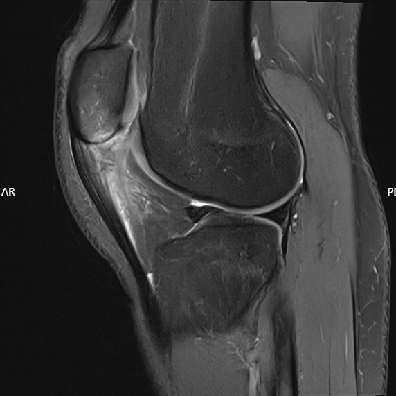
Dr. Gilmer's take:The exam for patellar tendinitis and its classic history are so common that I don't typically require an initial MRI. I will obtain an early MRI if there is frank weakness or the symptoms have been present for a very, very long time when the patient presents to me. Otherwise, we'll start with the conservative measures, and if these fail to improve, obtain an MRI looking for telltale signs
- Thickening of the tendon
- Intrasubstance signal or fluid within the tendon fibers themselves
- Whether there is any signal in the bone itself
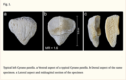
Interestingly, some different shapes or morphologies of patellas (where there is a longer extra-articular or non-cartilage surface of the patella and a shorter portion which is covered by cartilage) lend themselves to more frequent patellar tendonitis than other shapes.In those cases where the patient has a long tooth appearance to the distal pole of the patella, I will tend to be a little more aggressive early on, knowing that conservative measures are less likely to be effective.
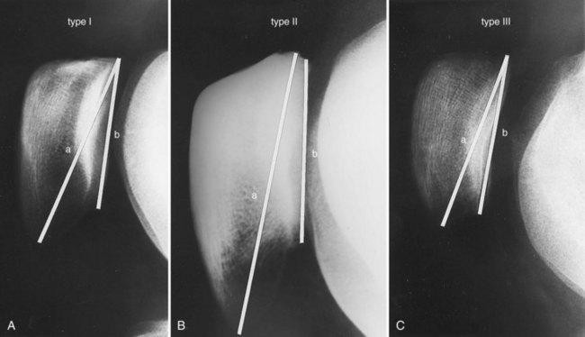
Figure 63-13 Different shapes of the patella in the sagittal plane according to Grelsamer and colleagues.43
Patellar shape is described by the morphology ratio, that is, the ratio of patellar length to length of the articular surface.
A, A normal type I patella has a morphology ratio between 1.2 and 1.5.
B, Type II patella, with a morphology ratio greater than 1.5, that is, with a long inferior pole (Cyrano appearance).
C, Type III patella with a morphology ratio below 1.2, that is, with a short inferior pole.
4. Surgery for delamination.
When delamination is confirmed and nonoperative treatment has been exhausted, the diseased portion of the tendon is debrided arthroscopically, the delaminated fibers are repaired back down to the patella with a single suture anchor, and a microfracture of the inferior pole of the patella is performed at the same time [3], [8]. The microfracture exposes bleeding cancellous bone, which is meant to stimulate healing at the bone-tendon interface being repaired, a principle similar to the bony decortication described in other arthroscopic patellar debridement techniques [8]. Published series of arthroscopic debridement for this condition report success in roughly 90% of patients, with return to sport around 3 to 5 months [3], [8]. Open reconstruction with tendon augmentation is reserved for more extensive detachment, generally greater than 50% of the tendon's width [7].
Surgical treatment is individualized. If there is just thickening of the tendon, a small incision can be made centrally, and the peritenon is cut, and the tendon is debrided internally. This is typically approached both externally and arthroscopically from beneath.
If there is a long inferior pole of the patella, this will be deburred with a bur or shaver arthroscopically to remove an area which can deflect the shape of the tendon and cause recurrence. If there is significant delamination, I will place a suture anchor. This does not change the recovery.
Ultimately, I have allowed patients to weight bear as tolerated and range of motion as tolerated in order to restore their function sooner, and have not seen any negative implications from this more aggressive rehabilitation pattern.
Post-operative Care
The microfracture site needs time to form a stable clot, and therefore patients are kept toe-touch or touch-down weight bearing in a brace locked in extension for the first 2 to 4 weeks, then gradually advanced toward full weight bearing by around 6 to 8 weeks. Range of motion is also protected early, generally limited to 0 to 90 degrees of flexion for the first several weeks before progressing further. Quadriceps activation exercises are started almost immediately to limit muscle atrophy, with more dynamic strengthening and sport-specific drills added once full weight bearing and motion are achieved. Return to jumping and cutting sports is typically allowed around 4 to 6 months, in line with the return-to-sport timelines reported after this procedure [3], [8].
Dr. Gilmer's take: As noted above, I have gradually moved away from long periods of bracing, as I find that early motion and stimulation result in better healing of the tendon, and there are some biomechanical studies to support this. On a more practical level, patients feel better and recover more quickly. Because the integrity of the tendon is intact, I'm less concerned about a need for a prolonged period of immobilization and find this to be counterproductive in many cases.
References
- P. Malliaras, J. Cook, C. Purdam, and E. Rio, “Patellar Tendinopathy: Clinical Diagnosis, Load Management, and Advice for Challenging Case Presentations,” Journal of Orthopaedic & Sports Physical Therapy, vol. 45, no. 11, pp. 887–898, Nov. 2015, doi: 10.2519/jospt.2015.5987.
- J. L. Cook and C. R. Purdam, “Is tendon pathology a continuum? A pathology model to explain the clinical presentation of load-induced tendinopathy,” Br J Sports Med, vol. 43, no. 6, pp. 409–416, Jun. 2009, doi: 10.1136/bjsm.2008.051193.
- J. P. van der List and C. C. Kaeding, “Arthroscopic Debridement of Proximal Patellar Tendinopathy in Athletes,” Video J Sports Med, vol. 6, no. 1, p. 26350254251359670, 2026, doi: 10.1177/26350254251359670.
- J. L. Cook, K. M. Khan, P. R. Harcourt, M. Grant, D. A. Young, and S. F. Bonar, “A cross sectional study of 100 athletes with jumper’s knee managed conservatively and surgically. The Victorian Institute of Sport Tendon Study Group.,” Br J Sports Med, vol. 31, no. 4, pp. 332–336, Dec. 1997, doi: 10.1136/bjsm.31.4.332.
- N. Maffulli, F. Oliva, M. Loppini, A. Del Buono, R. Aicale, and J. King, “The Royal London Test for the clinical diagnosis of patellar tendinopathy,” 2017, doi: 10.11138/mltj/2017.7.2.315.
- J. L. Dragoo, A. S. Wasterlain, H. J. Braun, and K. T. Nead, “Platelet-rich plasma as a treatment for patellar tendinopathy: a double-blind, randomized controlled trial,” Am J Sports Med, vol. 42, no. 3, pp. 610–618, Mar. 2014, doi: 10.1177/0363546513518416.
- J. S. Everhart et al., “Treatment Options for Patellar Tendinopathy: A Systematic Review,” Arthroscopy, vol. 33, no. 4, pp. 861–872, Apr. 2017, doi: 10.1016/j.arthro.2016.11.007.
- A. Amirhekmat, H. Stepanyan, K. Callan, R. WilliamsIII, and D. Wang, “Arthroscopic Patellar Tendon Debridement and Distal Pole Osteoplasty for Recalcitrant Patellar Tendinopathy,” Video Journal of Sports Medicine, vol. 2, no. 1, p. 26350254211054855, Jan. 2022, doi: 10.1177/26350254211054855.
- A. Scott et al., “Platelet-Rich Plasma for Patellar Tendinopathy: A Randomized Controlled Trial of Leukocyte-Rich PRP or Leukocyte-Poor PRP Versus Saline,” Am J Sports Med, vol. 47, no. 7, pp. 1654–1661, Jun. 2019, doi: 10.1177/0363546519837954.